
Make Your Research
More Accurate
More Efficient
Cloud-based AI platform for brain image analysis

Neurophet SegPlus is
an AI-powered platform to analyze brain image for research use.
Automated Process
Fully automated process enables image analysis without complicated preparation.
Quantitative analysis
Provides quantified analysis values and analyzed images suitable for each type of analysis.
Trial Version
Trial version of Neurophet SegPlus can be provided only for non-commercial and non-medical purposes, 3 times a day.
* Please consult us if you want to use analysis results obtained from the trial version for publication or presentation.Features
.png?id=b268ed35-15d3-44bc-868e-24f0f6a88b26&table=block&spaceId=9453ab34-9a3e-45a8-a6b2-ec7f1cefbd7f&width=2000&userId=&cache=v2)
AI-powered brain image analysis
The analysis can be completed within minutes using advanced AI engine and the accuracy is superior.
Automated process and integration
Fully automated AI engine analyzes images and integrates the result data into personal image processing pipeline.
Convenience and accessibility
Easily analyzes and manages in the web environment regardless of individual’s infrastructure environment and the result data can be accessed from anywhere at any time.
Analysis type
Neurophet SegPlus provides various AI analysis technologies for brain research. Experience efficient image analysis technology.
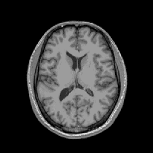
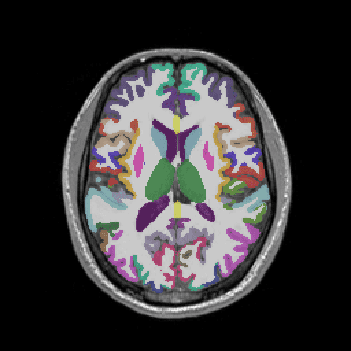
AI analysis that automatically segments T1-w brain MRI into multiple regions of interests(ROI).
Provides quantified volume information with normative percentile based on our own database.
Key feature

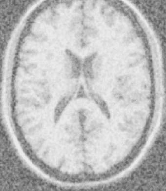
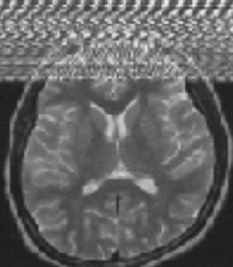
AI analysis that automatically segments T2-FLAIR brain MRI into white matter hyperintensities(WMH).
Provides quantified volume information of WMH.
Key feature
Trial version
Anyone can access to Trial version after signing up.
Experience the features of Neurophet SegPlus.
Security & Protection
Neurophet SegPlus prioritizes protecting private medical information..

All PHI is automatically
de-identified
Protected health information(PHI) defined by HIPPA regulations is automatically anonymized.
Easy to use, Strong security
Automatically encrypt the medical data according to HIPAA and safely transmit customer data through security protocols.
Workflow
Fast and simple workflow optimized for research
Provides optimized workflow according to various analysis types.
Step 1
Select the MRI image to analyze.
All PHI is automatically anonymized.

Step 2
Analysis results can be viewed directly on the web browser.
You can view the results from anywhere at any time.

Step 3
Download detailed analysis results and apply to your study.
The result can be integrated into your research pipeline.

1. Psychiatry investigation, Development of Random Forest Algorithm Based Prediction Model of Alzheimer’s Disease Using Neurodegeneration Pattern
2. Brain Sciences, Split-Attention U-Net: A fully convolutional network for robust multi-label segmentation from brain MRI
It depends on the type of analysis, it will be completed within 5 minutes in case of Brain Volumetry Analysis.
There may be a delay to start analysis depending on the number of jobs in the queue.
You will be notified by e-mail you registered on account when the analysis starts.
It automatically de-identifies all PHI in image according to HIPAA regulations.
If you want stronger security, please de-identify it yourself or convert it to NIfTI format.
De-identifies the following DICOM tags.
- [0008,0018] SOP Instance UID
- [0008,0020] Study Date
- [0008,0021] Series Date
- [0008,0022] Acquisition Date
- [0008,0023] Content Date
- [0008,0030] Study Time
- [0008,0031] Series Time
- [0008,0032] Acquisition Time
- [0008,0033] Content Time
- [0008,0050] AccessionNumber
- [0008,0080] InstitutionName
- [0008,1010] StationName
- [0008,1030] Study Description
- [0008,103e] Series Description
- [0010,0010] Patient's Name
- [0010,0020] Patient ID
- [0010,0030] Patient's Birth Date
- [0010,0040] Patient's Sex
- [0010,1010] Patient's Age
- [0018,1030] Protocol Name
- [0020,000d] Study Instance UID
- [0020,000e] Series Instance UID
- [0020,0010] Study ID
Neurophet SegPlus enables stable analysis of images taken according to the recommended parameters.
Please check that there is no reason for failure such as deviation from the recommended MRI parameters or artifacts.
If the same problem occurs in the images with the recommended parameters, please contact support@neurophet.com or use the inquiry.
Only analytical numerical values are available to download currently.
If it is for research purpose, please send us content on your paper or presentation material through inquiry or e-mail to support@neurophet.com.
The 'Trial version' to experience Neurophet SegPlus provides 3 times analysis per day.
If it is for research purpose, please send us content on your paper or presentation material through inquiry or e-mail to support@neurophet.com.
After an internal review, we will provide you extended license for your study through a written agreement.
Please send us content on your paper or presentation material through inquiry or e-mail to support@neurophet.com.
After an internal review, we will provide you extended license for your study through a written agreement.
Neurophet actively supports neuroscience and medical research.
Volumetry analysis
Type |
Structural MRI(3D T1-weighted) |
|---|---|
Slice thickness |
1 mm |
Pixel spacing |
0.8mm < spacing < 1.2mm |
No. of slices |
192 |
FOV |
256 x 256 |
Plane |
SAG |
* The image must contain entire brain area.
WMH lesion analysis
Type |
2D T2 FLAIR |
|---|---|
Slice thickness |
5 mm |
No. of slices |
20 ↑ |
Fov |
220 x 200 |
Plane |
Transversal |
* Slice thickness + slice gap ≤7mm
* Fat suppression images recommended.
Neurophet defined the analysis exception data as follows.
Following images may fail to be analyzed or may provide inaccurate analysis results even if they are analyzed.
Noise
Zipper
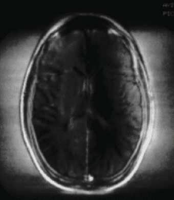
RF overflow
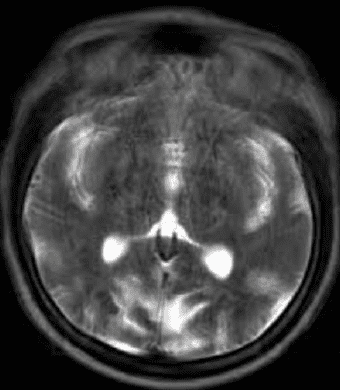
Motion
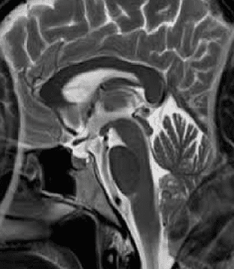
Fov error

Enlarged ventricle
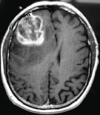
Lesion
(Brain Tumor)
Lesion (Stroke)
General
Artifacts
Brain structural changes
Experience Neurophet SegPlus
Request Trial